Research
Our understanding of mammalian tissue regeneration has been significantly reshaped in recent years by the discovery that many tissues, such as the heart, brain, spinal cord, and tendons, can mount a regenerative repair response at the neonatal stage independent of stem or progenitor cells, despite being previously recognized as non-regenerate in adults. Systemic and tissue-level changes occur following birth that include the transition from a relatively hypoxic in utero environment to ambient oxygen levels, a metabolic substrate shift from maternally supplied glucose to fatty acids in breast milk, and postnatal tissue growth and maturation. During this period, the immune system shifts the primary site of hematopoiesis from the fetal liver to the bone marrow and adapts from a protected in utero environment to the microbial world outside. The loss of neonatal regenerative capacity over time is likely an adaptation to these increasing physical, immunological, and energetic demands of adult life. However, how these changes converge at cellular and molecular levels that ultimately impedes tissue regeneration is not well understood.
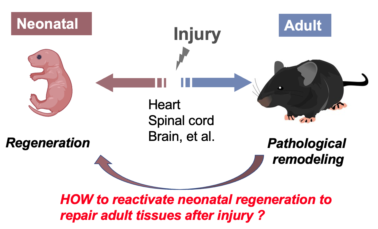
My lab uses the neonatal mouse heart regeneration model to study the underlying principles of neonatal tissue regeneration and its blockade in later life. Our research focuses on the tissue intrinsic regulation and systemic control of heart regeneration at the levels of individual cells and tissue organization. By integrating transcriptomics, proteomics, and metabolic profiling with tissue whole-mount imaging and genetic screens, we aim to understand the interplay among different cell types and the spatiotemporal regulation of genes during the 3-dimentional tissue reconstruction. Our research seeks to inform the development of regenerative strategies and enhance our understanding of cellular plasticity and stress responses that differ between neonates and adults despite sharing the same genome. Our research explores the intersections of regenerative biology, neonatology, immunology, and systems biology.
The origin and identity of regenerative cardiomyocytes.
Cardiomyocytes (CM) are the heart muscle cells integral for generating contractile force. Restoration of lost CMs after injury plays a vital role in driving the heart's functional recovery. While it is known that newly formed CMs during neonatal heart regeneration are derived from preexisting CMs, our understanding remains limited regarding the source of these generative CMs, their specific location within the heart, and their interactive behaviors during regeneration. We have identified a novel CM population (named CM4) that is unique to newborn mice and exhibits regeneration features following injury (Cui, et al., Dev Cell, 2020).
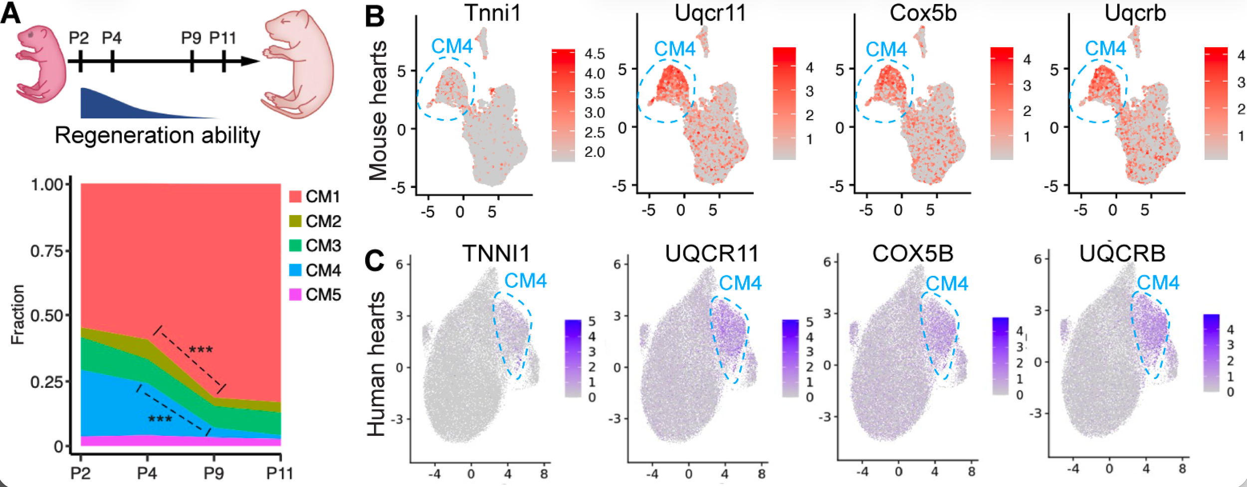
Our preliminary data show that these CM4 cells share a gene expression signature with zebrafish regenerative CMs and a CM population enriched in human infants, indicating an evolutionarily conserved regenerative program. Using a dual recombinase lineage tracing system, we found that these CM4 cells lineage contribute to the healed myocardium and are required for heart regeneration, as shown by cell ablation studies. Our current research is focuses on (1) Determine the genetic programs underlying the regenerative competence in these cells, (2) Understand injury-induced signals that stimulate their regenerative responses, (3) Understand mechanisms underlying the loss of this regenerative program postnatally over time, and (4) Identify upstream transcription factors capable of reactivate this regenerative state in adult heart.
Regeneration-conducive injury microenvironment
Regeneration requires coordinated responses across diverse cell types to restore tissue structure and function. Signaling networks are essential for cellular communication, yet how they are integrated within the injury microenvironment to produce coherent tissue-level repair remains poorly understood. To address this, we conducted spatial transcriptomic profiling, which identified the injury border zone as a signaling niche that coordinates repair in regenerating neonatal mouse hearts and identified 14 ligand–receptor pairs that may facilitate such regenerative responses, including previously implicated OSM and SLIT signaling.
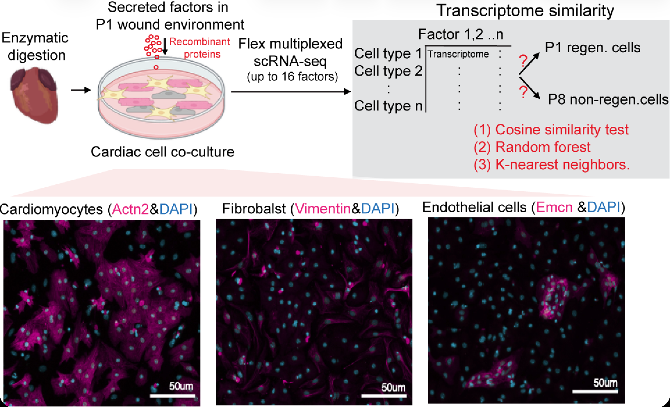
We developed SCREEM-seq (Single-Cell REgenerative Environmental Mapping by sequencing), a platform for cell type-resolved functional mapping of signaling ligands in a cardiac tissue-derived culture system, to systematically interrogate the functions of these neonatal-enriched signaling factors. Using this method, we found that many regeneration-associated ligands elicit broad transcriptional responses across cardiac cell populations but drive distinct, cell type-specific regenerative phenotypes, demonstrating that tissue repair emerges from coordinated, multi-lineage responses to a shared signaling landscape.
Our current research focuses on: (1) delineating cell type-specific regenerative functions using machine learning-guided transcriptomic phenotypic analysis; (2) identifying combinations of signaling factors that can promote complementary regenerative responses by functioning through different cell types; and (3) demonstrating the therapeutic potential of reprogramming tissue repair through targeted modulation of microenvironmental signals.
Regenerative proteome remodeling
Much of our current understanding of regeneration has been derived from transcriptomic studies; however, proteins are the primary effectors of cellular function, executing the metabolic and structural remodeling necessary to drive diverse regenerative processes, including dedifferentiation, proliferation, and redifferentiation. Consequently, successful regeneration depends on the ability of cells to dynamically remodel their proteome in response to injury. This remodeling requires a balance between the synthesis of new proteins to support growth and repair and the degradation of damaged or misfolded proteins to maintain protein quality control. These processes collectively define protein homeostasis, or proteostasis, which is essential for cellular adaptation during injury and disease. Importantly, these processes can occur independently of changes in transcript abundance, such as translational regulation of pre-existing mRNAs and selective degradation, enabling rapid proteome remodeling following injury. Understanding how cells coordinate dynamic proteome remodeling during tissue regeneration could identify new mechanistic insights and strategies to promote better tissue repair.
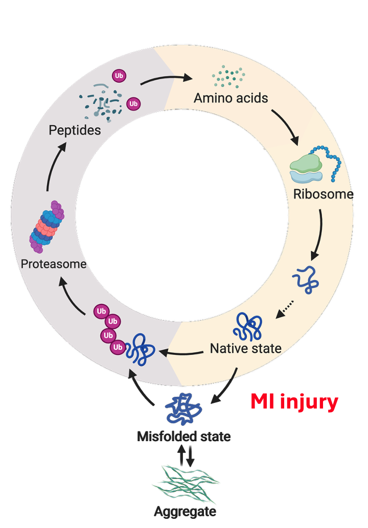
Our lab identified that neonatal regenerative hearts have improved ability to maintain proteostasis, accompanied by coordinated and simultaneous increase in both protein synthesis and degradation. We show that mTORC1 signaling is activated in regenerative cardiomyocytes at the injury border zone after injury and is responsible for the injury-induced increase of protein synthesis as well as protein degradation, thus is a central regulator for the regenerative proteome remodeling. Our current research is focused on (1) Identify the mechanism through which mTORC1 regulates protein degradation, (2) Understand how mTORC1 is activated at the border zone cardiomyocytes during regeneration, (3) Identify whether specific proteins are selectively translated and degraded and their potential regenerative functions.
Identify molecular contributors and barriers to heart regeneration using high-throughput functional in vivo screens
The limited regenerative capacity of the adult heart reflects both the loss of pro-regenerative programs and the acquisition of molecular barriers that restrict cardiomyocyte plasticity and proliferation. Identifying the key regulators underlying these changes is essential for understanding why regenerative capacity is lost after birth and for discovering molecular targets that could restore regenerative potential to the adult heart.
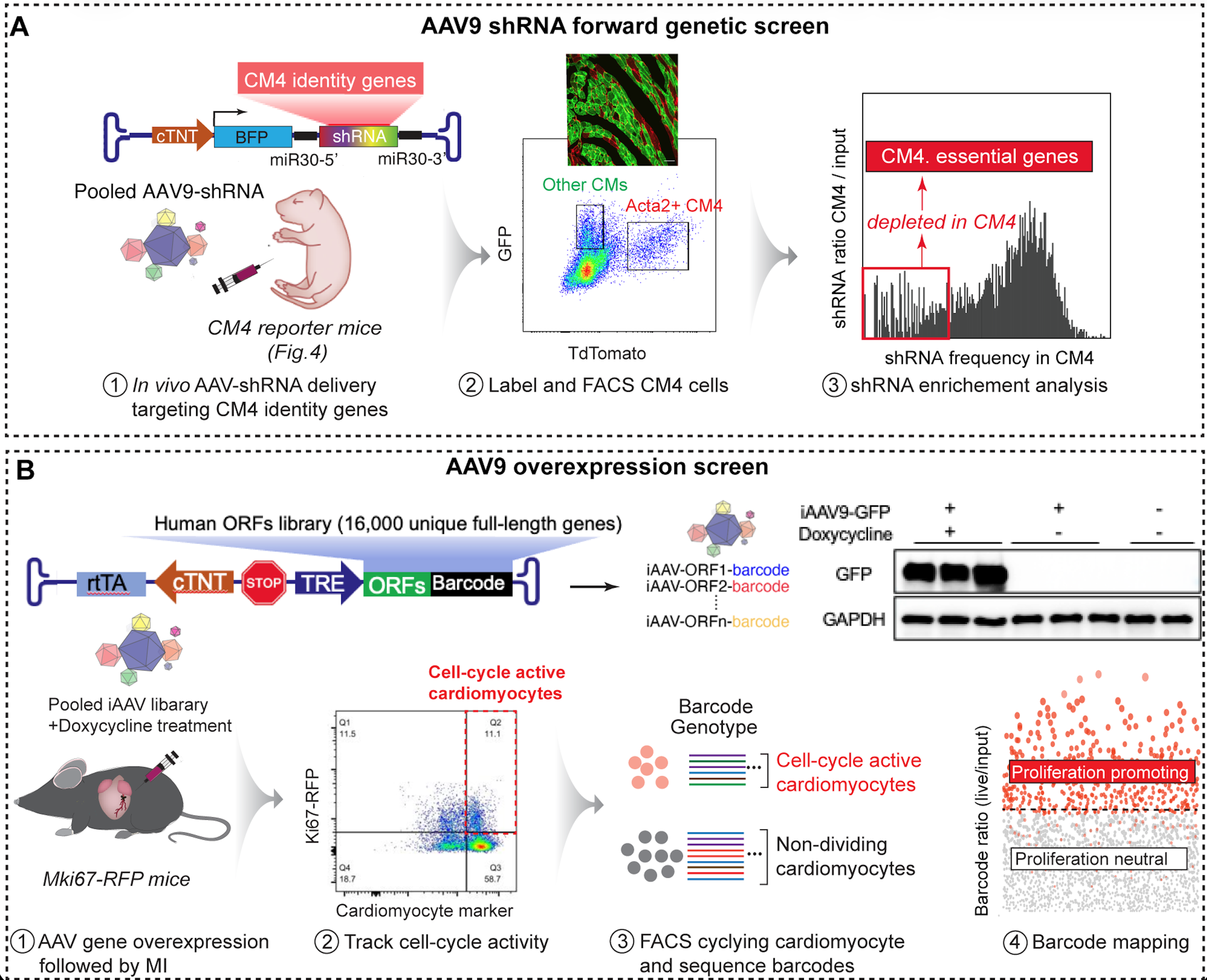
We integrate single-cell transcriptomic and chromatin accessibility analyses with network analysis and machine learning to identify candidate regulators of heart regeneration. We then use high-throughput AAV-based in vivo loss- and gain-of-function screens—including a genome-scale human ORF library and targeted shRNA libraries—to systematically test their regenerative functions in the heart. Together, these approaches aim to identify genes and pathways that promote regenerative cardiomyocyte states or remove barriers to regeneration, providing new targets and strategies for therapeutic heart repair.
Harnessing neonatal immunosuppressive mechanisms for tissue repair
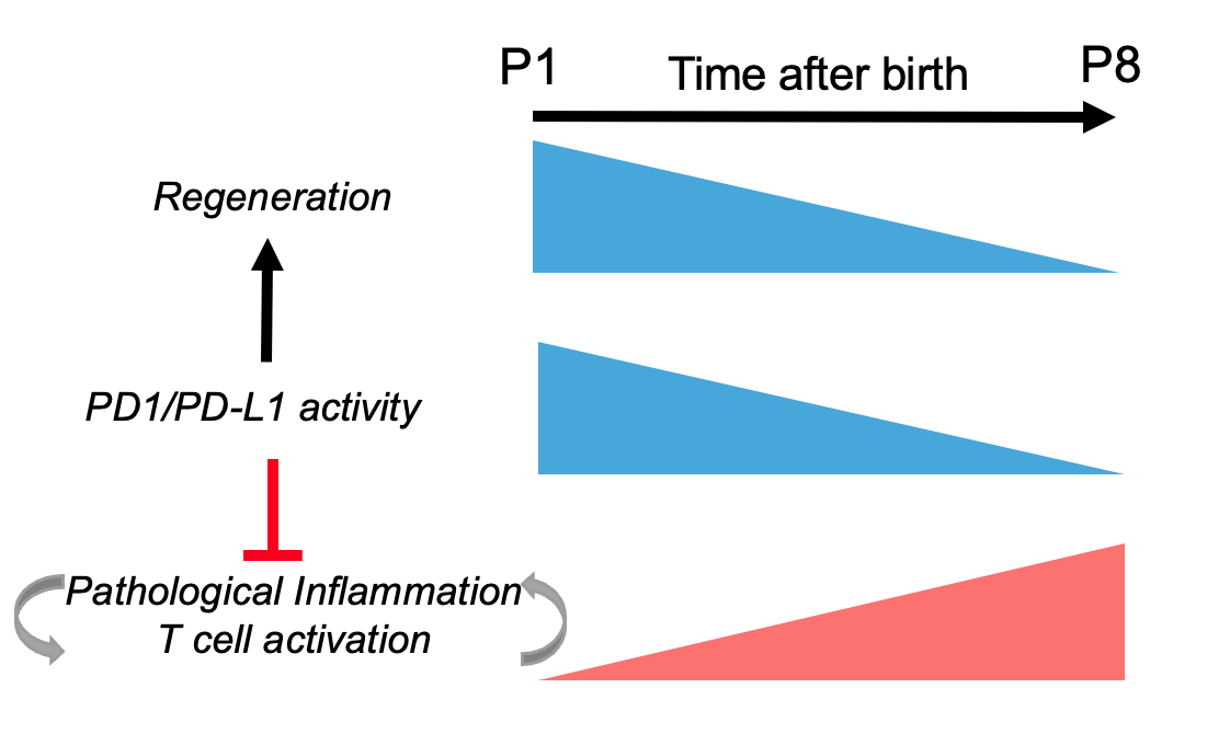
The ability of multiple tissues to regenerate in neonates suggests there are shared regenerative mechanisms regulated at the systemic level. The recruitment of circulating immune cells from blood and spleen to the injury site is among the first events after tissue damage. Previous research has demonstrated that the neonatal heart elicits a distinct immune response after injury that promotes cardiomyocyte proliferation and angiogenesis and is rapidly resolved as the heart regenerates. In a published work, we showed that the rapid resolution of the neonatal immune response is critical for heart regeneration and is partly regulated by the PD1/PD-L1 immune checkpoint pathway. While this work shed light on the previously unrecognized physiological role of this pathway that has been the most extensively studied in cancers, it also raised many questions that we are interested to further study. Does the heightened systemic PD1/PD-L1 pathway activity in neonates also contribute to regeneration in other tissues? Are there additional immune checkpoint pathways involved in neonatal tissue regeneration? Could enhancing these immunosuppressive mechanisms prolong the neonatal regenerative window?
Role of adaptive stress response in tissue regeneration and repair

The specific mechanisms by which cells in regenerative tissues adapt to stress conditions caused by injury during the regeneration process are still not fully understood. In two of our published work focusing the stress adaptive factor Nrf1 ( Cui et al, 2021; Kankanamge et al, 2026), we demonstrated that regenerative cardiomyocytes not only undergo cell-cycle activation but also upregulate cell survival pathways, including antioxidant response as well as proteasome and autophagy -mediated protein quality control. Our findings suggest a potential co-regulation between cardioprotection and heart regeneration. Traditionally, cardioprotection and heart regeneration were believed to involve distinct mechanisms. However, protecting cardiomyocytes from injury or disease stimuli is an essential prerequisite for any meaningful regenerative response. Our research focuses on exploring the unique stress adaptive mechanisms of neonatal regenerative cardiomyocytes and investigating how these mechanisms can be harnessed to enhance the reparative potential of adult hearts. We aim to demonstrate the therapeutic potential of these approaches using AAV (adeno-associated virus) and modified RNA molecules.


Cui Lab
Department of Cardiology
Boston Children’s Hospital
Department of Genetics
Harvard Medical School